
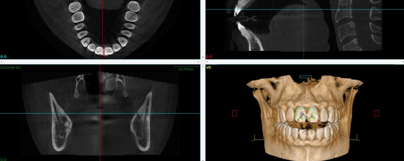
A cone beam CT creates a 3D view and cross-sections of the target area. The information it provides is critical for a number of situations. Our office utilizes this technology to aid in performing complex root canals or to search for the source of pain, infection or identified potential cracks. CBCT gives us the information we need about your condition before a dental procedure. It is not required for every case and is only taken when necessary.
Dental cone beam CT emits less radiation and provides a more complete picture than CT and CAT scans. Hospital CTs work by taking a series of parallel X-ray images of the head, from top to bottom. That leaves gaps between each image, which have to be filled by the computer’s educated guesses, and that many X-rays come with a lot of radiation.
A cone beam CT circles the head, which overlaps each image or slice, leaving no gaps. The radiation is also much weaker. The most radiation hits the area of interest, which is where the images overlap to construct the 3D model. This is how CBCT is able to provide a more complete image with less radiation exposure.
A CBCT scan gives much more detailed information than a regular dental X-ray, while using much less radiation than a medical CT scan. The small amount of radiation is considered very low risk and is often safer than missing an important diagnosis.
Because of this, CBCT scans are only recommended when regular X-rays do not provide enough information for an accurate diagnosis.
When needed, a CBCT scan — together with a review by a Maxillofacial Radiology Specialist — helps Dr. Grossman, Dr. Geisler, and Dr. Abitbol diagnose your condition more accurately and plan the best treatment for you.
For more information about 3D cone beam imaging, call us at (905) 668-6747 or send us an email.